CureSureMedico
|
EN

Interventional Bronchoscopy

Pulmonology

payments

Starting from

$1,500

Up to $8,000 depending on centre

schedule

Typical duration

3–10 days

Including pre-operative work-up

arrow_downwardlocal_hospital

Partner hospitals

1

Vetted for outcome data & accreditation

Dr. Rodina Ainasoa

Content reviewed for medical accuracy by: Dr. Rodina Ainasoa · Medical Content Writer & Health Communications

Overview

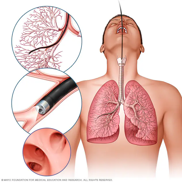
Advanced bronchoscopic procedures including endobronchial ultrasound (EBUS) for lymph node staging, rigid bronchoscopy for airway obstruction, metallic stenting, and bronchial thermoplasty — performed by specialist interventional pulmonologists. These minimally invasive techniques diagnose and treat complex airway and lung diseases without open surgery.

Your case is matched to a centre on the basis of clinical outcome data, not marketing. We coordinate every step — from pre-operative work-up to post-treatment follow-up — with a dedicated care coordinator.

What's included

Interventional pulmonologist consultation and pre-procedure CT review
Bronchoscopic procedure under sedation or general anaesthesia
Histopathology and cytology of biopsy specimens
Post-procedure monitoring and same-day or overnight stay
Written procedure report and specimen results in English
Medical coordinator support and teleconsultation for result review

Interventional Bronchoscopy

Coordinated from

$1,500

or message us on WhatsApp

No commitment required. Your data is never shared with third parties.

Conventional Electromagnetic Navigation vs. Robotic-Assisted Bronchoscopy

Reaching small nodules in the outer regions of the lung for biopsy can be done with conventional electromagnetic navigation bronchoscopy (ENB) or with a newer robotic-assisted bronchoscopy platform. Here is how the two approaches compare.

ParameterConventional Electromagnetic Navigation BronchoscopyRobotic-Assisted Bronchoscopy
How the bronchoscope is guidedA thin catheter is steered through the airways using an electromagnetic map built from your pre-procedure CT scan.A robotic system with a steerable catheter is controlled by the physician from a console, offering finer articulation and steadier positioning in the smallest peripheral airways.
Published diagnostic yieldRoughly 60-70% for peripheral pulmonary lesions, per peer-reviewed pulmonology literature.Published series report higher yields, up to roughly 88% with some robotic platforms, though results vary by platform, nodule size, and location — this remains an area of active study.
AvailabilityWidely available at hospitals with interventional pulmonology services.Offered at a smaller, though growing, number of specialist centres with the robotic platform and a trained interventional pulmonologist.
Safety profileA well-established, favourable safety profile, with pneumothorax and bleeding reported as uncommon complications.A similarly favourable safety profile in published comparative studies, with complication rates comparable to conventional navigation.
Typical procedure settingPerformed under moderate sedation or general anaesthesia, often combined with other diagnostic steps such as EBUS lymph node sampling in the same session.Also performed under moderate sedation or general anaesthesia; the choice between conventional and robotic-assisted navigation is made by the interventional pulmonologist based on nodule characteristics and platform availability.

The Procedure: Step by Step

The steps below outline a typical navigational bronchoscopy procedure for sampling a lung nodule. Your interventional pulmonologist will confirm the exact plan based on your imaging and the nodule's location.

1

Pre-procedure imaging and planning

A recent CT scan of the chest is used to build a virtual roadmap of your airways and pinpoint the location of the nodule or lesion to be sampled, planning the safest route to reach it.

2

Sedation and bronchoscope insertion

Under moderate sedation or general anaesthesia, a thin bronchoscope is passed through the mouth or nose into the airway, and a steerable catheter is guided toward the target using the electromagnetic or robotic navigation system.

3

Tissue sampling and, if needed, mediastinal staging

Once the catheter reaches the target area, specialised tools are used to collect tissue or cell samples for biopsy; endobronchial ultrasound (EBUS) may be performed in the same session to sample nearby lymph nodes if cancer staging is required.

4

Recovery and specimen review

You are monitored in a recovery area as sedation wears off, with a chest X-ray typically performed to check for pneumothorax before discharge; biopsy specimens are sent to pathology, with results usually available within several days.

Recovery Timeline

Recovery Timeline
Same day

Most patients are observed for several hours after the procedure and discharged the same day once sedation has fully worn off and a follow-up chest X-ray shows no complications, per Mayo Clinic and Cleveland Clinic.

First 24-48 hours

A mild sore throat, hoarseness, or minor blood-tinged sputum from the biopsy site is common and typically resolves on its own; you should avoid driving or operating machinery until sedation has fully cleared.

First few days

Most people return to normal, non-strenuous activity within a day or two; your care team will advise if follow-up imaging is needed to monitor the biopsy site or confirm nodule characteristics.

Follow-up

Biopsy and, where performed, EBUS lymph node results are reviewed with your pulmonologist or oncology team to plan next steps; if a sample is inconclusive, a repeat procedure or an alternative biopsy approach may be recommended.

Source: American College of Chest Physicians (CHEST); American Thoracic Society; Mayo Clinic; Cleveland Clinic. Individual recovery varies and is guided by your treating interventional pulmonologist.

Known Risks & Limitations

  • Pneumothorax (a partially collapsed lung) is a recognised risk of navigational bronchoscopy, particularly when sampling nodules near the lung's outer edge; most cases are minor and resolve with observation, though some require a temporary chest tube, per CHEST and peer-reviewed pulmonology literature.
  • Bleeding at the biopsy site can occur and is usually minor and self-limiting, though more significant bleeding is an uncommon but recognised complication requiring closer monitoring or intervention.
  • Diagnostic yield varies meaningfully by nodule size, location, and the presence of a bronchus leading directly to the lesion (the 'bronchus sign'); smaller or more peripheral nodules are harder to sample accurately regardless of the navigation technology used.
  • Sedation and, where general anaesthesia is used, its associated risks apply, including reactions to anaesthetic agents and, in patients with significant lung disease, a higher anaesthesia risk that your team will assess beforehand.
  • If the tissue sample obtained is inconclusive or insufficient for diagnosis, a repeat bronchoscopy, a CT-guided needle biopsy, or a surgical biopsy may be needed to reach a definitive diagnosis.

Source: American College of Chest Physicians (CHEST); American Thoracic Society; Mayo Clinic; Cleveland Clinic; peer-reviewed pulmonology literature. No procedure is without risk; your treating interventional pulmonologist will review your individual case.

The CureSureMedico Care Pathway

Remote case review

Share your chest CT or PET-CT imaging, nodule characteristics, and any prior biopsy attempts with our coordination team, who forward your case to an interventional pulmonologist for an initial suitability review.

Specialist and platform matching

We match you with an accredited hospital and interventional pulmonology team experienced in your specific procedure, including access to a robotic-assisted platform where clinically appropriate and available.

Cost estimate and travel planning

Once your case is reviewed, we issue a consolidated cost estimate covering the procedure, sedation or anaesthesia, and pathology, and help plan a trip length that allows time for recovery and result review before you fly home.

Procedure and monitoring

You attend your bronchoscopy procedure and are monitored afterward with a follow-up chest X-ray before discharge, in line with standard post-procedure safety checks.

Result review and continuity of care

Biopsy and staging results are reviewed with your treating team before you depart, and your full procedure report and pathology results are shared with your home-country physician for ongoing care.

Treatment approaches

Standard and robotic-assisted Interventional Bronchoscopy

Standard Approach

Conventional Electromagnetic Navigation Bronchoscopy (ENB)

$1,500

310 days

Robotic-Assisted Option

Robotic-Assisted Navigational Bronchoscopy

$3,000

310 days

Comparing the two approaches

StandardRobotic-Assisted
ProcedureConventional Electromagnetic Navigation Bronchoscopy (ENB)Robotic-Assisted Navigational Bronchoscopy
TechnologyConventional surgical instrumentsRobotic-assisted surgical system with surgeon oversight
AvailabilityHospital-dependentSelected hospitals with robotic capability
SpecialistProcedure specialistSpecialist with relevant robotic experience
Clinical suitabilityCase-dependentCase-dependent
Tariff$1,500–$8,000$3,000–$12,000
Duration3–10 days3–10 days

Eligibility for robotic-assisted treatment

  • A peripheral pulmonary nodule or lesion identified on CT imaging as a candidate for bronchoscopic biopsy, rather than one clearly better suited to CT-guided needle biopsy or surgical excision
  • No anatomical factors, such as extreme peripheral location without a clear bronchus leading to the lesion, that would substantially limit reachability regardless of navigation technology
  • Interventional pulmonology review of imaging confirming candidacy and, where relevant, the need for concurrent mediastinal staging
  • Availability of the specific robotic bronchoscopy platform and an interventional pulmonologist experienced in robotic navigation at your selected hospital — offered at fewer centres than conventional electromagnetic navigation bronchoscopy

Risks and limitations of robotic-assisted surgery

  • Robotic assistance does not eliminate the risks of navigational bronchoscopy, including pneumothorax, bleeding, and the possibility of an inconclusive sample
  • The interventional pulmonologist remains responsible for the procedure; robotic guidance is an aid to catheter stability and reach, not a substitute for clinical judgement
  • Published studies report a favourable diagnostic-yield trend for robotic-assisted platforms versus conventional electromagnetic navigation for peripheral nodules, though results vary across studies, platforms, and nodule characteristics, and comparative evidence is still accumulating
  • Availability depends on the individual hospital and the treating specialist's experience with the specific robotic platform

Tariff structure

Standard procedure$1,500$8,000
Robotic-assisted technology fee+$1,500$4,000
Total estimated robotic-assisted tariff$3,000$12,000

Why can robotic-assisted procedures cost more?

Robotic-assisted procedures rely on additional technology and equipment beyond the standard operating platform and clinical team. This additional technology component is generally not included in standard case pricing.

Is robotic-assisted surgery always recommended?

No. Robotic-assisted surgery is not appropriate for every procedure or patient. Availability and suitability are determined by the treating specialist, based on your imaging, clinical condition, and the treating hospital's capabilities.

Global cost comparison — Interventional Bronchoscopy

Average coordinated costs across our partner centres. Final pricing depends on clinical complexity and chosen hospital.

CountryAvg. costvs. cheapest
🇮🇳

India

New Delhi · Chennai · Bengaluru · Hyderabad · Mumbai · Kochi

Best value
$2,000
chevron_right
🇹🇷

Turkey

Istanbul

$5,000
chevron_right

Costs are indicative estimates and vary by procedure specifics, hospital, and clinical complexity. Request a personalised estimate for your specific case.

Partner hospitals

1 centres
View all hospitals
Amrita Hospital

Amrita Hospital

Faridabad, India

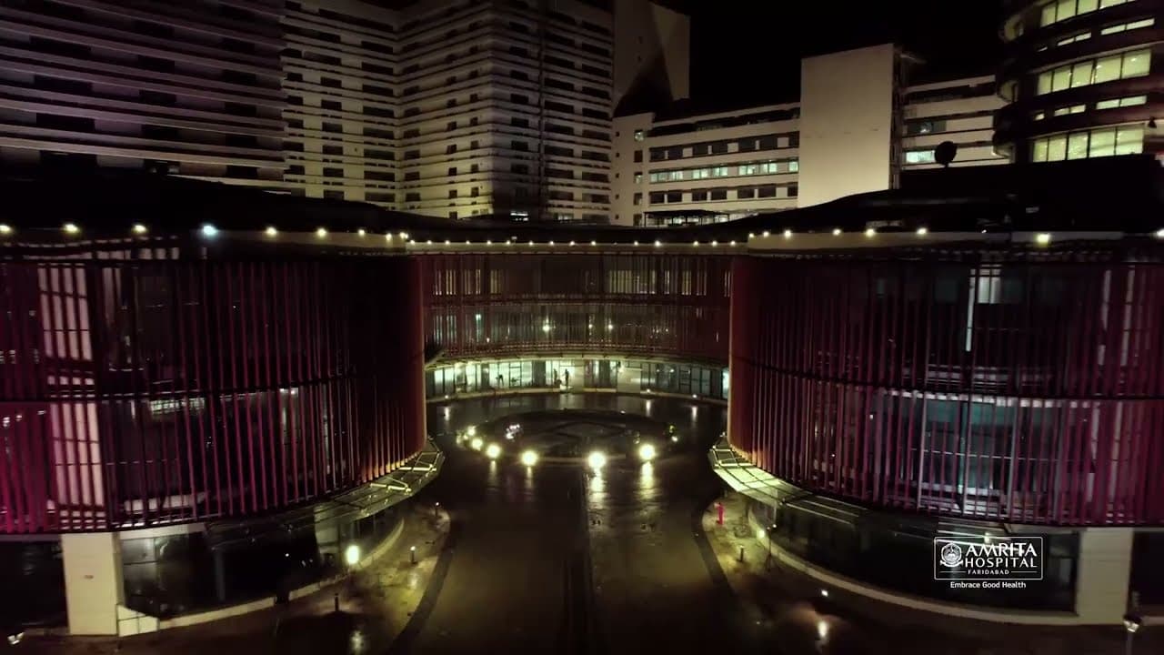
Asia's largest private hospital — 2,600 beds, 64 operation theatres, 81 specialties on a 130-acre campus in Delhi NCR. NABH & NABL accredited. Centres of excellence in oncology, cardiac surgery, BMT, organ transplantation, neurosciences, and IVF.

NABHNABL

800+

Specialists

2,600+

Beds

View Profile

Frequently asked questions

EBUS-TBNA (endobronchial ultrasound with transbronchial needle aspiration) is performed entirely through the mouth via a bronchoscope equipped with an ultrasound probe, allowing real-time visualisation and sampling of mediastinal and hilar lymph nodes without any skin incision or radiation exposure from fluoroscopy. It is the preferred first-line method for mediastinal staging of lung cancer, with a diagnostic yield of over 90% for accessible nodes, and carries a lower complication risk than CT-guided biopsy for central lesions.

No fees. No commitment.

Our guidance is completely free

We are compensated by our partner hospitals — never by patients. You get independent clinical matching, cost transparency, and end-to-end coordination at no cost to you.

Message us on WhatsApp