Aortic stenosis is among the most widespread and serious structural heart valve conditions encountered in adult cardiology. Over time, calcium deposits build up on the aortic valve, causing it to narrow and stiffen. Restricted in its function, the heart must work under immense pressure to pump oxygenated blood to the body, leading to symptoms such as shortness of breath, chest pain, dizziness, fatigue, and fainting. Left untreated, severe symptomatic aortic stenosis significantly compromises quality of life and carries a high risk of heart failure and mortality.
For decades, open-heart Surgical Aortic Valve Replacement (SAVR) was the sole definitive treatment for aortic stenosis. However, open-heart surgery requires a sternotomy (opening of the chest bone), cardiopulmonary bypass (stopping the heart), and a prolonged recovery period—making it high-risk or unfeasible for elderly patients or individuals with co-existing medical conditions.
Transcatheter Aortic Valve Implantation (TAVI), also referred to as Transcatheter Aortic Valve Replacement (TAVR), represents a major breakthrough in interventional cardiology. TAVI allows heart specialists to replace a damaged aortic valve via a catheter inserted through a small vascular access point, eliminating the need for open-heart surgery.
Expert Video Overview & Clinical Insights
For international patients across Sub-Saharan Africa, accessing specialized cardiovascular interventions requires careful clinical evaluation, multidisciplinary review, and well-coordinated travel logistics. Planning this journey ensures that candidates are thoroughly screened prior to departure, guaranteeing seamless transfer to leading cardiac centers with minimal delay and maximum safety.
To help patients and families understand the technical nuances and multidisciplinary teamwork involved in transcatheter valve replacement, watch the medical video below featuring the team at Centre Cardio-Thoracique de Monaco (CCM Channel) demonstrating the TAVI procedure and high-tech hybrid imaging:
Source: "Implantation percutanée d'une prothèse valvulaire aortique (TAVI)" — CCM Channel.
Key medical insights highlighted by the clinical specialists in the video include:
Watch the cardiovascular specialists at the Centre Cardio-Thoracique de Monaco explain the TAVI procedure in detail above.
Multidisciplinary Heart Team Collaboration: "The TAVI procedure is conducted within a state-of-the-art hybrid operating room where interventional cardiologists, cardiac surgeons, vascular surgeons, and cardiac anesthesiologists work in synchronized unison." — Centre Cardio-Thoracique de Monaco Clinical Team
Anchoring Within Native Structures: "Unlike open surgery where the diseased valve is excised, TAVI anchors the new artificial valve directly inside the calcified native valve leaflets, utilizing the native calcium structure as a secure foundation." — Centre Cardio-Thoracique de Monaco Clinical Team
Precision Hybrid Imaging: "Modern hybrid operating rooms utilize robotic C-arms and 3D image fusion—combining pre-procedural CT scans with live fluoroscopy—to navigate catheters and position the valve with millimeter precision." — Centre Cardio-Thoracique de Monaco Clinical Team
What Is TAVI / TAVR?
Transcatheter Aortic Valve Implantation (TAVI)—commonly referred to as TAVR in North America—is a minimally invasive, catheter-based procedure designed to replace a severely narrowed aortic valve.
Anatomical Mechanism and Access Routes — The aortic valve sits between the left ventricle (the heart's main pumping chamber) and the aorta (the main blood vessel delivering blood to the body). During TAVI, a collapsed artificial valve is navigated through the arterial system using specialized delivery catheters:
Transfemoral Access (Primary Route): In over 90% of cases, access is achieved percutaneously via a small puncture in the femoral artery in the groin. The catheter is guided up through the iliac artery and aorta into the heart.
Alternative Access Routes: If a patient has severely diseased, narrow, or calcified leg arteries, alternative routes are utilized—such as transsubclavian (under the collarbone), transaortic (direct access via the upper chest), or transapical (via a small incision in the chest wall directly into the tip of the heart).
Valve Technology & Anchoring Mechanism
Unlike surgical valve replacement, which cuts away the calcified native valve leaflets and sutures a new valve in place, TAVI anchors the prosthetic valve inside the native valve.
The native calcium deposits serve as an anchoring frame for the expansion of the new valve. The prosthetic valves typically consist of tissue leaflets (such as bovine or porcine pericardium) mounted on a collapsible cobalt-chromium or nitinol metal stent frame. The frame is deployed inside the aortic annulus using either balloon inflation or self-expanding technology.
How a Heart Team Evaluates TAVI Candidacy
TAVI may be considered for selected patients with severe, symptomatic aortic valve stenosis or failing structural bioprosthetic valves (Valve-in-Valve procedure). Whether TAVI is an appropriate option—and which valve replacement approach is used—depends on a combination of factors, including age, valve and vascular anatomy, surgical risk, life expectancy, and other medical conditions. This determination is made by a multidisciplinary Heart Team of cardiologists and cardiac surgeons, not through patient self-assessment. Symptoms that commonly prompt this evaluation include exertional dyspnea (shortness of breath during normal daily activities), angina or chest pressure during physical exertion, presyncope or syncope (dizziness, lightheadedness, or sudden fainting), chronic fatigue and reduced exercise tolerance, and signs of congestive heart failure (fluid retention, leg swelling).
Examples of Factors a Heart Team Assesses — The criteria below illustrate the kinds of clinical assessments a Heart Team uses to evaluate candidacy; they are examples of that process, not a checklist patients can apply to determine their own eligibility. TAVI was initially approved for patients considered inoperable or at high surgical risk, and its use has since expanded:
High & Prohibitive Surgical Risk: The Heart Team may consider TAVI for patients aged 80 years and above, or those with severe comorbidities (such as chronic kidney disease, prior coronary artery bypass grafting, frailty, or porcelain aorta), among other factors weighed during evaluation.
Intermediate & Low Surgical Risk: Following thorough Heart Team evaluation, TAVI may also be considered for select intermediate-risk and lower-risk older adults.
Treatment Comparison: TAVI vs. Traditional Surgery
Understanding the differences between TAVI and conventional Surgical Aortic Valve Replacement (SAVR) helps patients and families make informed treatment choices:
| Comparative Parameter | Transcatheter Aortic Valve Implantation (TAVI / TAVR) | Surgical Aortic Valve Replacement (SAVR) |
|---|---|---|
| Invasiveness | Minimally invasive; percutaneous needle puncture in the groin. | Invasive open-heart surgery; requires median sternotomy. |
| Cardiopulmonary Bypass | Not required; performed on a beating heart. | Required; heart is stopped and blood redirected to a heart-lung machine. |
| Anesthesia Type | Local anesthesia with conscious sedation or light general anesthesia. | Full general anesthesia with endotracheal intubation. |
| Native Valve Handling | Native calcified valve remains in place; new valve expands inside it. | Diseased calcified valve is excised and sutured manually. |
| Hospital Stay | Typically 1 to 3 days. | Typically 5 to 10 days (including dedicated ICU stay). |
| Recovery Period | Rapid return to mobility (within 24–48 hours); full recovery in 1–2 weeks. | Extended recovery period (6 to 12 weeks for bone healing). |
Diagnostic Workup: Key Pre-Procedure Evaluations
Prior to undergoing TAVI, a comprehensive diagnostic protocol is mandatory to measure the aortic root's dimensions, select the correct prosthetic valve size, and verify arterial access pathways.
ECG-Gated Cardiac Computed Tomography (Cardiac CT): The primary planning tool for TAVI. Accurately measures the aortic valve annulus area, perimeter, coronary ostia height, and degree of calcification. Evaluates the entire vascular tree (iliac and femoral arteries) to verify catheter access diameter.
Transthoracic & Transesophageal Echocardiography (TTE / TEE): Measures pressure gradients across the aortic valve, aortic valve area, and left ventricular ejection fraction.
Coronary Angiography (Cardiac Catheterization): Checks for concurrent coronary artery disease. If significant blockages exist, stenting (PCI) may be performed prior to or during TAVI.
Comprehensive Laboratory Workup: Includes complete blood count, renal function testing (creatinine, eGFR to ensure contrast dye clearance), and coagulation profiles.
Intraoperative Overview
Preparation: The patient is placed in a hybrid catheterization suite equipped with advanced fluoroscopy and 3D fusion imaging.
Vascular Access: The clinical team establishes percutaneous access via the femoral artery in the groin.
Crossing the Valve: A specialized guidewire is carefully navigated across the narrowed aortic valve into the left ventricle.
Rapid Pacing & Valve Deployment: To temporarily drop cardiac output and stabilize positioning, rapid ventricular pacing is initiated. The prosthetic valve is precisely expanded inside the native valve.
Verification: Angiography and echocardiography confirm valve opening, coronary flow clearance, and check for paravalvular leaks.
Closure: Vascular closure devices seal the femoral access site without requiring surgical sutures in the groin.
Post-Procedure Recovery & Monitoring
Immediate Post-Op: Patients are monitored in a cardiac telemetry unit for 12 to 24 hours.
ECG Monitoring: Continuous monitoring tracks the heart's electrical conduction system to rule out heart block.
Discharge: Most patients are ambulatory within 24 hours and discharged within 2 to 4 days.
Clinical Benefits
Immediate Symptom Relief: Patients experience rapid improvement in shortness of breath, chest pressure, and energy levels.
No Open Chest Incision: Avoids sternotomy complications and minimizes post-operative pain.
Rapid Recovery: Enables older or frail patients to regain mobility and independence within days.
No Heart-Lung Machine: Reduces systemic inflammatory stress associated with cardiopulmonary bypass.
Possible Risks and Complications
Conduction Disturbances (Pacemaker Need): Expansion of the valve frame near the heart's electrical pathways may require a permanent pacemaker in a small percentage of patients.
Vascular Complications: Bleeding, hematoma, or injury at the femoral access site.
Paravalvular Leak: Minor blood leakage around the outer frame of the prosthetic valve.
Stroke Risk: Micro-calcification debris can dislodge during positioning; embolic protection filters are frequently used to minimize risk.
Expert Insights Summary
As emphasized by the clinical team at the Centre Cardio-Thoracique de Monaco:
On Procedural Precision: "Success in transcatheter valve implantation relies on meticulous pre-procedural CT planning, ensuring that the prothesis fits the annulus perfectly while preserving coronary artery flow."
On Patient Selection: "TAVI offers a transformative, minimally invasive alternative for patients who face high surgical risks or who desire a rapid return to an active, independent life."
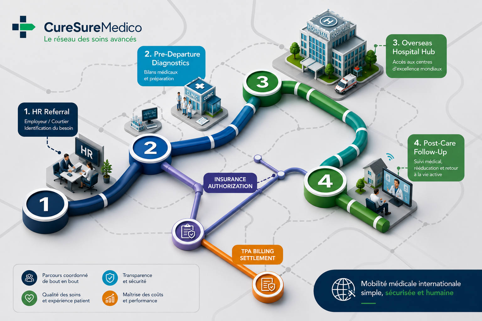
International Medical Care with CureSureMedico
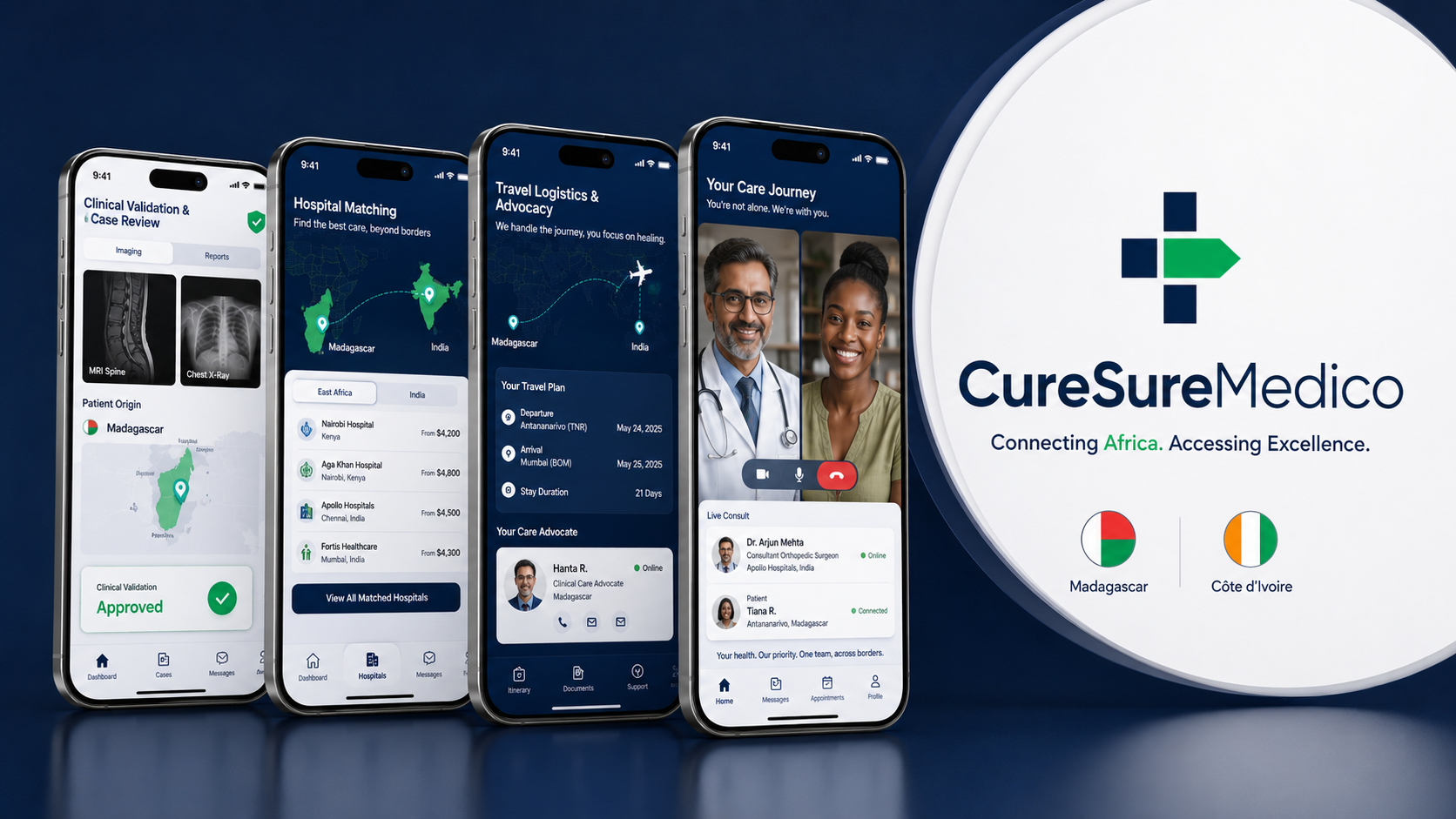
For patients residing across Sub-Saharan Africa who require advanced structural heart procedures like TAVI, organizing medical travel requires seamless clinical oversight and logistical support. CureSureMedico provides a complete, transparent, and patient-centered framework:
Remote Diagnostic Review & Second Opinions: CureSureMedico collects your local echocardiograms, cardiac CT scans, and medical reports. Our medical advisory panel coordinates pre-travel reviews with international interventional cardiologists to confirm procedural suitability.
Transparent Flat-Fee Model: CureSureMedico operates with direct hospital billing and transparent flat-fee case management—ensuring no hidden markups or commissions on medical treatment.
Pre-Travel Diagnostic Verification: Our team ensures all high-resolution cardiac CT scans meet strict TAVI sizing protocols before you fly, eliminating unnecessary travel expenses or unexpected delays.
Comprehensive Logistics Facilitation: We manage medical visa support letters, priority hospital scheduling, airport transfers, hotel accommodations for family members, and dedicated bedside assistant services.
Continuity of Care Back Home: Following your return home, CureSureMedico facilitates post-discharge tele-consultations, digital record sharing, and coordination with your local treating cardiologist in Africa.
Considering TAVI Treatment?
If you or a loved one has been diagnosed with severe aortic stenosis, CureSureMedico can help you access an independent cardiology review and coordinate a seamless treatment pathway with leading international TAVI centers.
Start with a clinical review — contact a CureSureMedico advisor today to evaluate your case and discuss transcatheter aortic valve treatment options.
Frequently Asked Questions
How long does a TAVI prosthetic valve last?
Modern bioprosthetic TAVI valves (typically made from bovine or porcine pericardium) demonstrate excellent durability, with clinical studies showing structural valve performance comparable to surgical valves over 5 to 10+ years.
Is TAVI performed under general anesthesia?
Not always. While TAVI can be performed under general anesthesia, many leading international centers perform transfemoral TAVI under local anesthesia with conscious sedation, allowing faster post-procedural recovery.
Will I need to take blood thinners after TAVI?
Most patients are prescribed antiplatelet therapy (such as aspirin and/or clopidogrel) for a few months post-procedure to prevent blood clots on the valve frame. Your cardiologist will tailor the prescription based on your individual medical history.
How soon can international patients fly back home after TAVI?
Most patients are up and walking within 24 hours. International patients are typically advised to stay near the treating hospital for 5 to 7 days post-discharge for a routine follow-up echocardiogram before taking their flight home.